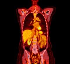
July 29, 2014 — New study data presented at the Alzheimer’s Association Intl. Conference 2014 (AAIC) showed that a positive [18F]flutemetamol positron emission tomography (PET) scan for brain amyloid was a highly significant predictor of progression from amnestic Mild Cognitive Impairment (aMCI) to probable Alzheimer’s disease (pAD). Those with positive [18F]flutemetamol scans were approximately 2.5 times more likely to convert to pAD than those with negative scans. The ability of positive [18F]flutemetamol PET images to identify aMCI patients at higher risk of progressing to AD could potentially allow for better patient evaluation and management.
“These findings demonstrate the potential role of [18F]flutemetamol in stratifying those patients at higher risk of developing Alzheimer’s disease, beyond its use as a diagnostic tool,” said David Wolk, M.D., assistant director, Penn Memory Center and lead investigator of the study. “In addition to providing patients with potentially important prognostic information about their likelihood of developing dementia, identifying high risk patients could help guide physicians’ recommendations for patient monitoring, care plans and use diagnostic resources. These are exciting results, but we need further research to fully understand how this might be used in clinical practice.”
In the study, 232 participants with MCI, a diagnosis characterized by cognitive deficits not severe enough to impact daily functioning and thus not meeting the definition of dementia, received a [18F]flutemetamol injection and underwent brain scans. Images were read visually by five independent blinded readers and classified as [18F]flutemetamol negative or positive for the presence of brain amyloid. Subjects were then assessed clinically onsite every six months until progression to pAD (determined by an independent Clinical Adjudication Committee) or for three years, whichever occurred first.
A Cox proportional hazards model was used to estimate hazard ratios (HR) for progression to pAD. The median HR for an amyloid positive image was 2.58 and was >2.5 for each of 4 readers. The HR was highly significant for each individual reader. The findings also demonstrated that patient age was highly significant in predicting conversion to pAD for each reader (median HR 1.05). As expected, those patients with late-stage aMCI were more likely to progress to pAD than those with early-stage aMCI (median HR 2.199).
In October 2013, [18F]flutemetamol received approval from the U.S. Food and Drug Administration (FDA), where it is marketed as Vizamyl for PET imaging of the brain to estimate beta amyloid neuritic plaque density in adult patients with cognitive impairment who are being evaluated for Alzheimer’s disease (AD) or other causes of cognitive decline. Vizamyl is for diagnostic use only and should be used in conjunction with a clinical evaluation. Vizamyl is not licensed in any market for estimating the risk of MCI progression to clinical AD.
In June 2014, [18F]flutemetamol received a positive opinion from the European Medicines Agency’s Committee for Medicinal Products for Human Use (CHMP), recommending the granting of a marketing authorization for PET imaging of beta amyloid neuritic plaque density in the brains of adult patients with cognitive impairment who are being evaluated for Alzheimer’s disease and other causes of cognitive impairment. It is not yet approved for use in Europe or Japan.
The results of this study were presented at AAIC as a late breaking poster entitled, “[18F]Flutemetamol Amyloid PET Imaging: Outcome of Phase III Study in Subjects with Amnestic Mild Cognitive Impairment after 3 Year Follow Up.” The study was sponsored by GE Healthcare.
For more information: newsroom.gehealthcare.com


 July 30, 2024


