July 28, 2014 — Santa Fe Imaging (SFI) has become the first facility in New Mexico and the entire mountain west region to offer 3-D automated whole breast ultrasound (ABUS) for the early detection of breast cancer in women with dense breast tissue. Breast density increases the risk of breast cancer and makes it more difficult to detect by traditional mammography alone.
According to the American College of Radiology (ACR), more than 40 percent of all U.S. women have dense breast tissue, and women in the highest dense-breast category established by the ACR are four to six times more likely to get breast cancer compared to women with low breast density.
Breast ultrasound uses sound waves to generate a picture of the breast tissue, and no compression is necessary. Santa Fe Imaging Radiologist Monica Saini, M.D., emphasized that breast ultrasound does not replace mammography, which is still the gold standard, but ABUS is a new and important screening supplement available to women.
“Adding 3-D automated breast ultrasound to mammography in dense-breasted women gives physicians a valuable new tool in the fight against breast cancer,” said Saini. “Physicians get a clearer evaluation of dense breast tissue, which can significantly increase the rate of cancer detection at an earlier, more treatable stage.”
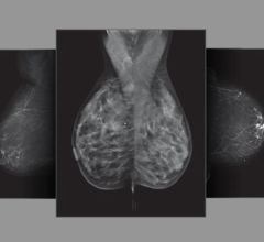
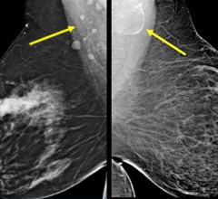
For women with less dense, fatty tissue, cancer shows up on mammography as white spots on a dark (fatty tissue) background. However, for women with more dense glandular tissue, cancer appears white on a white (dense tissue) background, making it difficult and sometimes impossible to identify. With 3-D automated whole breast ultrasound screening, cancer shows up black on a white background, making it much easier to identify.
Research and awareness of breast density is on the rise, and it has led to 17 states passing notification laws requiring that women be informed of their breast density if their mammogram comes back negative. New Mexico does not yet have such legislation, but Saini said SFI has taken a proactive approach to the issue by notifying women of breast density in the exam results letter and informing them of other breast imaging options.
“We reviewed this issue, and based on the evidence we believe it is important to provide dense breast notification to our patients,” said Saini.
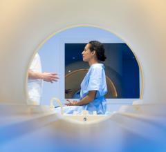
Radiologists have routinely reported breast density to patients’ referring physician for years; however, it is not typically part of the standard results letter that women receive if their mammogram is negative. Supplemental screening at SFI includes breast ultrasound and breast magnetic resonance imaging (MRI). When used in conjunction with mammography, such tests may find hidden cancer, leading to improved early detection.
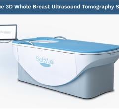
Desert Rose Women’s Center at SFI uses the Invenia Automated Breast Ultrasound technology, which is clinically proven to improve breast cancer detection by 35.7 percent compared to mammography alone. The technology is especially useful at detecting smaller and earlier stage cancers in women with dense breasts, but it is also useful in telling cysts from solid masses. Supplemental imaging with ABUS is covered by insurance for most dense-breasted patients.
For more information: www.santafeimaging.com


 July 29, 2024

