March 19, 2014 — Molecular breast imaging (MBI) Clinical Indications, Benefits and Challenges is the focus of a new CME credit course to be offered by ICPME. It is funded in part by Gamma Medica Inc. and in part by GE Healthcare. The one-hour online session will provide a thorough overview of MBI, which is a growing imaging technology for women with dense breasts and certain difficult-to-image cancers.
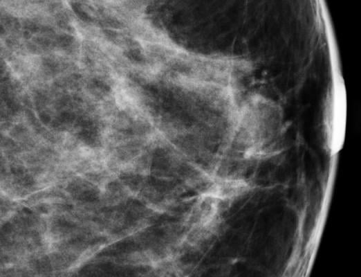
Research has shown that for women with dense breasts, MBI technology outperformed mammography in finding more cancers. This makes MBI, which has the ability to detect breast cancer lesions as small as 5 mm, a highly effective secondary diagnostic tool. This is particularly true for women with dense breast tissue and who have inconclusive results on their initial mammograms.
MBI’s ability to detect smaller lesions lies in its ability to use molecular functional imaging, allowing for breast abnormalities to be detected based on altered characteristics of the tissue. Functional imaging is important for distinguishing between a viable and nonviable mass because functional changes precede anatomical changes in breast tissue.
The certified pre-recorded session is presented by Stephen W. Phillips, M.D., FACR, medical director of Methodist Sugar Land Hospital Breast Center (MSLH), assistant professor of radiology at Weill Cornell Medical College, New York, and formerly assistant professor of radiology at Mayo Clinic. Since 2012, Phillips has played a role in MBI clinical research at MSLH, where he currently utilizes MBI as a contributor to the general health and well-being of women in the greater Texas area.
The course assesses the role of MBI as a complementary tool to mammography, describes patient indications for use, evaluates MBI benefits and challenges and covers the counseling of patients with dense breast tissue. It is accredited for 1.0 AMA PRA Category 1 credit and participants can register on the ICPME.
For more information: courses.icpme.us


 July 30, 2024


