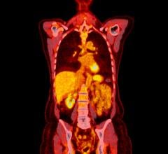
An FDDNP brain scan of an individual with mild cognitive impairment (MCI) illustrates the parietal and frontal regions of the brain (see arrows) that have significant relationships to cognitive function. The lateral temporal lobe was another significant region, but is not included in this brain section.
April 5, 2013 — UCLA researchers have used a brain-imaging tool and stroke risk assessment to identify signs of cognitive decline early on in individuals who don't yet show symptoms of dementia.
The current small study demonstrated that not only stroke risk, but also the burden of plaques and tangles, as measured by a UCLA brain scan, may influence cognitive decline.
The imaging tool used in the study was developed at UCLA and reveals early evidence of amyloid beta (plaques) and neurofibrillary tau (tangles) in the brain — the hallmarks of Alzheimer's disease.
The study, published in the April issue of the Journal of Alzheimer's Disease, demonstrates taking both stroke risk and the burden of plaques and tangles into account may offer a more powerful assessment of factors determining how people are doing now and will do in the future.
"The findings reinforce the importance of managing stroke risk factors to prevent cognitive decline even before clinical symptoms of dementia appear," said first author David Merrill, M.D., Ph.D., an assistant clinical professor of psychiatry and biobehavioral sciences at the Semel Institute for Neuroscience and Human Behavior at UCLA. “This is one of the first studies to examine both stroke risk and plaque and tangle levels in the brain in relation to cognitive decline before dementia has even set in.”
According to the researchers, the UCLA brain-imaging tool could prove useful in tracking cognitive decline over time and offer additional insight when used with other assessment tools.
For the study, the team assessed 75 people who were healthy or had mild cognitive impairment, a risk factor for the future development of Alzheimer's. The average age of the participants was 63.
The individuals underwent neuropsychological testing and physical assessments to calculate their stroke risk using the Framingham Stroke Risk Profile that examines age, gender, smoking status, systolic blood pressure, diabetes, atrial fibrillation (irregular heart rhythm), use of blood pressure medications and other factors.
Each participant was also injected with a chemical marker called FDDNP, which binds to deposits of amyloid beta plaques and neurofibrillary tau tangles in the brain. The researchers then used positron emission tomography (PET) to image the brains of the subjects — a method that enabled them to pinpoint where these abnormal proteins accumulate.
The study found that greater stroke risk was significantly related to lower performance in several cognitive areas, including language, attention, information-processing speed, memory, visual-spatial functioning (e.g., ability to read a map), problem-solving and verbal reasoning.
The researchers also observed that FDDNP binding levels in the brain correlated with participants' cognitive performance For example, volunteers who had greater difficulties with problem-solving and language displayed higher levels of the FDDNP marker in areas of their brain that control those cognitive activities.
"Our findings demonstrate that the effects of elevated vascular risk, along with evidence of plaques and tangles, is apparent early on, even before vascular damage has occurred or a diagnosis of dementia has been confirmed," said the study's senior author, Gary Small, M.D., director of the UCLA Longevity Center and a professor of psychiatry and biobehavioral sciences.
Researchers found that several individual factors in the stroke assessment stood out as predictors of decline in cognitive function, including age, systolic blood pressure and use of blood pressure–related medications.


 July 30, 2024


