Dr. James Craft of the GHSU Breast Health Center says the additional clarity the 3D images provide enables them to be more confident about their diagnoses.
Kellie Bedenbaugh, a registered breast imaging technologist at the Breast Health Center at Georgia Health Sciences Medical Center, is excited about the hospital’s recent installation of two tomosynthesis systems and what it means for her patients. “Tomosynthesis helps our radiologists see inside the breast much more thoroughly. The images acquired produce a high-quality digital mammogram along with images that the computer divides into multiple layers, which the radiologist can view one millimeter slice at a time,” explains Bedenbaugh.
The Breast Health Center is part of Georgia Health Sciences Health System, the clinical arm of Georgia Health Sciences University (GHSU). An academic health center, GHSU prides itself on its combination of research, technical expertise and patient care. The hospital installed two Hologic Selenia Dimensions digital mammography systems in February and added the Dimensions digital tomosynthesis capability three months later.
The Breast Health Center uses the tomosynthesis system for all mammograms, both screening and diagnostic, and is the first health system in Georgia and one of only a handful in the country to make tomosynthesis the standard for care in breast imaging.
“The usual screening population is asymptomatic,” states Dr. James Craft, a breast imager at the Center and Assistant Professor of Radiology at GHSU. “Seventy-six percent of women who develop breast cancer don’t have any risk factors. We can’t predict who will get breast cancer, and the tomosynthesis dose is less than it was when we were doing film studies a year ago, so we give everyone the best exam and the best care that we can.”
Using Tomosynthesis to See Through the Tissue
Tomosynthesis images are acquired with the breast held briefly in compression. A screening examination, which includes both a 3D tomosynthesis scan and a 2D digital mammogram, takes only seconds longer than a conventional 2D digital mammogram at a total exam dose within the current FDA guidelines for annual mammography screening.
The 3D scan results in multiple sequential high-resolution slices that provide clear separation of structures in the breast and their spatial relationship with the surrounding breast tissue. Dr. Craft says tomosynthesis helps enhance some of the architectural distortion, making it easier to see where calcifications are located. “You can truly see through the layers,” he says.
Two mammographers and four technologists at the Breast Health Center perform approximately 7,000 mammograms annually. “The major benefit of tomosynthesis,” explains Dr. Craft, “is being able to see more.” In just a few short months, Dr. Craft and his colleagues have already seen results. “We diagnosed an intraductal invasive cancer using the 3D images; we did not see the cancerous area in the 2D images at all.”
Separating Real Abnormalities from Possible Abnormalities
Dr. Craft says tomosynthesis allows radiologists to detect cancers much earlier, before they become invasive. “With tomosynthesis, we can separate densities that were previously hidden by surrounding tissue which could not be separated. Now, these densities are more obvious sooner, resulting in fewer callbacks, less anxiety and, most importantly, earlier detection.”
Recently, Bedenbaugh took images of a breast cancer survivor with fatty breasts. The 2D images showed one mass. But when Dr. Craft looked at the tomosynthesis images, he saw a second mass behind the nipple that would have been completely missed without the 3D tomosynthesis images.
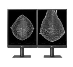
The Breast Health Center at Georgia Health Sciences Medical Center uses Hologic’s SecurView DX diagnostic workstation to read the mammograms. With digital mammography technology, the breast images are available immediately and the images are stored electronically. The SecurView workstation enables radiologists to manipulate the 2D and 3D images, enhance areas for a closer view, and compare images from other modalities side-by-side.
The breast imaging department also uses the Hologic ATEC vacuum-assisted breast biopsy device for use with stereotactic, MRI or ultrasound guidance, performing as many as three ultrasound biopsies a day. And the department recently added Hologic’s Aegis breast image analysis software for MRI.
The New Standard in Mammography
The University’s role is not just to provide the highest level of patient care, but also to train the next generation of healthcare professionals. To do that, GHSU ensures its staff has the most up-to-date knowledge and training. “Our residents are coming so far so quickly reading mammograms with tomosynthesis,” states Dr. Craft. “Tomosynthesis is helping them get to a level of confidence in what they are seeing much more quickly because they can look through the layers of the breast.
“I am very pleased with tomosynthesis,” concludes Dr. Craft. “The additional clarity that the 3D images provide enables us to be more confident about our diagnoses. It is a tremendous step forward – not only in what we see, but also in what it does for our patients.”
Views and opinions expressed herein by third parties are theirs alone and do not necessarily reflect those of Hologic.
Hologic, Aegis, ATEC, Dimensions, SecurView and Selenia are trademarks and/or registered trademarks of Hologic Inc. and/or its subsidiaries in the United States and/or other countries.
Case study supplied by Hologic Inc.


 July 29, 2024


