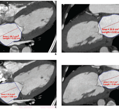
September 23, 2011 – Traditionally in computed tomography (CT), there has been an undesirable trade-off between image quality and low radiation dose levels. While high CT image quality often required greater patient exposure to diagnostic radiation, lower dose levels for the patient usually meant lower image clarity from higher noise and more artifacts.
Yet times are changing, and GE’s breakthrough CT technology – Veo – has taken the next step toward eliminating this trade-off.
After decades of CT image reconstruction using filtered back projection (FBP) and more recent advances in statistical raw data-based iterative reconstruction, such as GE’s powerful ASiR technology, Veo’s capabilities “change the rules” of CT imaging by applying revolutionary modeling techniques that open up possibilities for further dose reduction in clinical practice. GE Healthcare is coordinating a multi-center study to investigate this potential for Veo to deliver lower noise, increased resolution, improved low contrast detectability, fewer artifacts – and overall improved image clarity with lower dose than other forms of iterative and FBP reconstruction.
The CT industry’s first model-based iterative reconstruction (MBIR) technique, and already available on GE Discovery CT750 HD systems in Europe, Canada and regions of Asia, Veo may help physicians deliver accurate diagnoses by enabling profound CT image clarity at dramatically lower dose.
“The radiological community continues to demand maximum CT image clarity at optimized dose levels to help best serve our patients,” said William Shuman, M.D., professor and vice chairman, Department of Radiology at the University of Washington. “A superb image quality, sub-millisievert (mSv) CT exam has in many ways been seen as CT’s holy grail – making powerful new diagnostic tools like Veo all the more vital as we seek to minimize patient dose while maximizing diagnostic confidence. The Veo reconstruction option may also improve the image quality of low dose non-diagnostic CT images (filtered back projection), such that they become diagnostic.”
In fact, Veo has already opened the door to profound CT image clarity at yet unseen low-dose levels. Current Veo users in Europe report successful chest CTs done with an equivalent amount of medical radiation dose as a chest X-ray, or less than .1 millisievert (mSv).
“With a clinical chest CT at 0.05 mSv, we produced images where we could see and analyze pathology,” said Johan de Mey, professor and chair of the Radiology Department at University Hospital in Brussels, Belgium.
+In clinical practice, the use of Veo may reduce CT patient dose depending on the clinical task, patient size, anatomical location and clinical practice. A consultation with a radiologist and a physicist should be made to determine the appropriate dose to obtain diagnostic image quality for the particular clinical task." For more information:

 February 01, 2024
