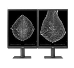
Breast imaging centers, like many other users of imaging systems, are jumping feet-first into digital technology. The conversion process from film to digital doesn’t have to be difficult, and digital systems offer many benefits to both users and patients, from improved workflow and reduced storage space requirements, to superior image quality.
Coleen Goulet, R.T.(R)(M) ARRT, is the diagnostic imaging manager at Sunnyside Community Hospital’s Breast Imaging Center in Sunnyside, Wash. Sunnyside’s center, which performs ultrasound-guided breast biopsies as well as mammograms, converted from filmscreen to digital mammography in January 2010, after finding a need to update its technology.
“We were seeing trends [toward digital] and thought it was the standard-of-care to be digital versus filmscreen,” Goulet said.
An important reason to transition to digital was that Goulet’s center saw women in younger age brackets developing breast cancer, and digital mammography is better at imaging the dense breast tissue many women in their 40s and younger have.
“Digital systems can see through dense tissue so much better,” Goulet said. “The radiologist can magnify, invert and move the image; before, it was just a piece of film with light shining through it and no opportunity for maneuvering.”
There were also storage concerns. Goulet said they were running out of space to store mammography images for a long period of time, as many other facilities do.
The Search Process
With these reasons in mind, Goulet and her team decided to look at digital mammography systems. They visited sites that hosted two systems they were considering, and Goulet said they were fortunate to be able to schedule both site visits on the same day.
“That helped a lot,” she said. “We were able to compare them when our memories of the machines were fresh, and that was very good for us.”
The team looked at factors such as image quality, speed of processed image (or how fast the image appears on-screen after being taken) and user-friendliness. They ultimately chose the Hologic Selenia digital mammography system.
“It seemed to be more user-friendly for us because we were acquainted with the system after having used Hologic in the past,” she said.
They picked the NovaRad NovaMG for their radiologist review station. Not only were the radiologists able to learn the system quickly because the center was already using NovaRad for its picture archiving and communication system (PACS), but also NovaMG is vendor-neutral, meaning the station can be used with any digital mammography vendor system.
“We’re a critical access hospital and we lease a lot of our equipment,” Goulet said. “In five years, if we decided to upgrade our mammography equipment, we would be stuck with the original vendor. But with NovaMG, we can choose any vendor.”
Simplifying the Transition
Goulet said it was essential to prepare staff ahead of time to ensure everyone was comfortable with the new system.
“Once we chose the mammography system, we got radiologists to do their digital training ahead of time, before we went live,” she said. “That was helpful in easing anxiety about the new system.”
Sunnyside’s center also made sure its information technology (IT) staff was in the loop about the installation so they could access DICOM information, as well as images from the PACS.
“It’s important to make sure the IT department is on board with installation, even pre-installation,” Goulet said.
Once Sunnyside’s imaging center installed its system, it became the first hospital in Washington’s Yakima Valley to have digital mammography. It wanted current and potential patients to know about the new technology. So the center informed the Valley’s newspaper and staff members were interviewed on television and radio programs about the new system.
“Our plan was to increase the number of patients who came here, and we did that, by about 10 percent,” Goulet said.
Film versus digital
The advantages to digital mammography are numerous, said Goulet. The TV spot showed clips of digital images side-by-side with a film image to show the difference between the two.
“Even without a trained eye, you can see the difference in image quality,” Goulet said. “It’s like looking at a picture on a digital camera versus a Polaroid.”
In addition to image quality, techs no longer have to process the images in a dark room; they appear on a monitor 20 seconds after being taken. Goulet said this has decreased the time required per mammogram from about 20 minutes to about 10. So the workflow has improved significantly.
There also are storage benefits, Goulet said. Instead of being kept in a physical storage facility, images are kept on a PACS server. NovaRad backs up the images nightly and keeps copies on its server in Utah in case something happened to Sunnyside’s server.
“For the hard copies of films, there’s no backup [for disaster recovery],” Goulet said. “If we need the films, we have to go get them.”
Future of the industry
One trend that Sunnyside’s center is seeing is growing use of breast magnetic resonance imaging (MRI). The center does not do it in-house, but sends patients to another larger facility.
“We’re seeing more MRI done than a few years ago, probably because of improvements in scanners and how well they can see masses [in breasts],” Goulet said.
Breast tomosynthesis is also a new industry development. Tomosynthesis is a 3-D imaging technology that involves acquiring images of a stationary compressed breast at multiple angles during a short scan. It is better at imaging dense breast tissue.
Sunnyside’s imaging center does not do tomosynthesis at this time, but Goulet said its machine is capable of doing it in the future.
“We wanted to make sure we would be upgradeable,” she said. “We had heard a little about it when we were looking at systems, but it wasn’t FDA-approved at the time.”
Goulet said she believes this is the next big step for breast imaging centers, but not for five to 10 years.
“I think the trend now is for 100 percent of facilities to go completely digital,” Goulet said. “The next thing after doing that is tomosynthesis.”


 July 29, 2024


