The medical imaging market is poised for growth, and among the areas showing great promise are hybrid systems, especially positron emission tomography/computed tomography (PET/CT), according to a new report, “Medical Imaging Markets,” by TriMark Publications. The report’s executive summary also notes the growing popularity of single photon emission computed tomography/computed tomography (SPECT/CT) in U.S. and global markets.
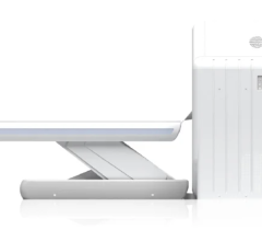
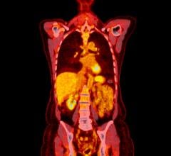
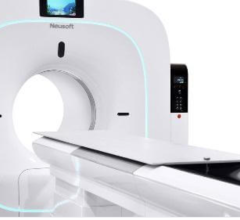
In PET/CT, functional information in the form of PET data and anatomic information in the form of CT data are acquired almost simultaneously, so the two information sets can be viewed and interpreted together, as the Society of Nuclear Medicine (SNM) explains it. The multi-detector CT apparatus and PET detectors are mounted in the same gantry, one immediately behind the other. For interpretation, the PET data is actually superimposed upon the CT data, SNM says.
According to a tutorial developed by the University of Virginia Health Sciences Center’s Department of Radiology, PET/CT images acquired simultaneously provide several advantages over PET scanning alone, where PET images would typically be correlated with CT images acquired at a different time. It says PET/CT is becoming the preferred choice over PET alone, and TriMark concurs, saying, “the future of dedicated PET is guaranteed no more.” Similarly, in an online overview of the molecular imaging market, SNM says, “The use of hybrid imaging equipment is on the rise. Today, virtually all PET units sold are hybrid devices.”
The SPECT/CT combination also is growing in use. The first system combined a dual-head gamma camera and an integrated X-ray transmission system mounted on the same gantry, according to SNM’s “Procedure Guideline for SPECT/CT Imaging 1.0.” More recent systems combine a multi-head gamma camera and multi-detector CT scanner side by side with a common imaging table.
“Although techniques for registration and fusion of images obtained from separate SPECT and CT scanners have been available for several years, the advantages of having SPECT and CT integrated into a single device have resulted in the development of this technology in the United States and elsewhere in the world,” SNM notes.
With these hybrid modalities garnering glowing reports, ITN asked some physicians who are using them for their thoughts about their pluses and minuses. They started with a long list of advantages.
Patrick F. Wojtylak (R) (N) CNMT PET, supervisor of nuclear medicine PET/CT at Case Medical Center, and Peter Faulhaber, M.D., associate professor at Case Western Reserve University in Cleveland, Ohio, have a Gemini TF 16 PET/CT system from Philips at their facility. Following are several benefits they have found using a hybrid system:
• They can perform both anatomical and functional imaging during the same patient visit, with no temporal lag waiting to retrieve magnetic resonance (MR) or CT and fuse; they also get better fusion alignment
• Attenuation correction is applied. This improves image quality which, in turn, increases referring physician satisfaction and, most importantly, patient diagnosis
• Low-dose CT decreases radiation exposure to the patient and can limit unnecessary radiology procedures
• It allows for better surgical planning; functional and anatomical combined helps surgeons plan the best tactic in addressing the patient’s issue. This can negate a hospital stay or shorten a stay and improve recovery time
• Hybrid imaging is the focal point for any radiation therapy planning and assessing response to treatment
• The ever-changing and dynamic field of hybrid imaging has a lot of potential for growth and with it, a facility is seen as state-of-the-art and very progressive
Joseph J. Busch, M.D., Diagnostic PET/CT of Chattanooga in Tennessee, says he feels there is a better utilization of technology with a hybrid imaging system. “There is no ‘dead time’ on high-dollar technology,” he says.
His facility has Biograph 16 HI-REZ and Biograph TruePoint 6 PET/CTs and a Symbia T6 SPECT/CT system from Siemens. He says with the hybrid systems, even if the scanner is down, a CT can be performed; plus nuclear studies may be performed without CT and vice-versa. He notes that his facility does use the hybrid systems to do general CT scans, CT-guided biopsies and surgery localization scans, besides hybrid imaging. A disadvantage is that if the CT is down, a PET scan can’t be performed, and if the CT is down on a SPECT system, the protocol must be changed to do cardiac work.
Ivan Huyghe, M.D., of the Universitair Ziekenhuis Antwerpen Nucleaire Geneeskunde in Edegem, Belgium, points to other advantages of hybrid systems. His facility has a Discovery NM/CT 670 SPECT/CT machine from GE Healthcare, and he sees the following benefits:
• Matching of CT and nuclear medicine (NM) are always perfect (unless the patient moves in between)
• It is easier to locate scintigraphic hotspots, especially in those exams with little or no anatomical references (131 Iodine scanning, Octreoscan, etc.)
• It shortens NM SPECT acquisition, because diagnosis can be made with poorer count statistic
• CT provides anatomical reference and attenuation correction
• He can obtain even better image quality with SPECT imaging using GE Healthcare’s Evolution reconstruction software technology, which allows the hospital to perform SPECT imaging on patients who are difficult to image, such as children, the elderly or other people who have a lot of discomfort while on the camera
• It provides the possibility of offering “one-stop shop” service to non-hospitalized patients for whom it is often difficult to travel to a hospital (e.g., cancer patients, diabetic foot patients, etc.)
• It is easy to determine the field of view of CT based on the NM persistence image
There are a couple of disadvantages, Huyghe says. “When you are not doing SPECT/CT, at least one of the modalities is standing by idle, and breakdown of one of the components often results in the other one not being usable either.”
And although it is not a problem at his hospital, Huyghe adds that in a facility where the machine is shared between two departments, such as radiology and NM, it could lead to conflicts.
Wojtylak and Faulhaber point to perhaps the biggest disadvantages, the price of the machine and lack of insurance reimbursement for hybrid imaging. But they do see the advantages they cited above as being a justification for the cost, adding that there is a return on investment in hybrid imaging in the form of increased referring physician satisfaction, which parallels the growth in patient volume.
Busch says Diagnostic PET/CT of Chattanooga has seen a return on investment for its hybrid systems. He says the cost justification is getting better utilization (more studies in less time) using less square footage, which also means fewer full-time employees.
Huyghe cites several points of justification for the expensive machine at his hospital. One is, “the ability of doing diagnostic CT with the same quality as a stand-alone dedicated CT system, while keeping the ability to do low-dose CT as well,” he says. “Because of this, at least in Belgium, we can get reimbursed by social security for this CT, which is not the case for low-dose CT. In this way, it was possible to have a co-financing agreement with the radiology department for our Discovery. It would not be the case for a Hawkeye-like system.”
He says that the machine’s ASiR (adaptive statistical iterative reconstruction) low-dose reconstruction technology is a must for his department, “because our radiology department has ASiR as well. We cannot justify doing the same diagnostic CT procedures with a radiation burden that would be higher on the Discovery system than on the Brightspeed 64 of the radiology department. ASiR makes it easier for physicians (who are nowadays under increasing pressure from the health department to lower radiation doses to patients) to send patients to our Discovery system instead of some other systems in neighboring hospitals.”
“Since the radiology department was involved in making the tender for this SPECT/CT system, to them the CT specs were very important,” he adds. “The ability to have iterative reconstruction and a platform like BrightSpeed that was still under active development were essential.”
Improved image quality is also a justification for the higher cost, according to Huyghe. “Having a fast CT will lead to shorten exam time and lower possibility of motion artifact,” he says. “Our powerful Volumetrix MI suite is a great tool for reading those images, due to the intuitive user interface, the ability of fast volume reorientation, 3-D rendering (which makes useful images for physician-patient discussion) and fusion capabilities with other DICOM sets, such as MRI, contrast-enhanced CT, etc.”
Huyghe says that determining whether his hospital has gotten a return on investment from the hybrid system is difficult to judge at this time because it was a fairly recent acqusition. “Although the Discovery was installed in July 2010, the department was not connected to the picture archiving and communications system (PACS) until December. Prior to that, it was impractical to perform diagnostic CT because of transfer problems to radiology. Also, ASiR has only been installed for a few months now, so we are just building up to the normal rate of SPECT plus diagnostic CT. Because of the delay in PACS and ASiR, we are only now starting to advertise the machine’s capabilities to our referring physicians, with increasing success.”


 July 30, 2024


