High-Quality X-Ray Images with Crisp Contrast Resolution

The PaxScan 1313 offers a 13-by-13- cm imaging area, produces up to 30 images per second, and is optimized for use in orthopedic imaging and semiconductor inspection systems.
Many orthopedic imaging systems currently have six-inch image intensifiers, and Paxscan 1313 is designed to replace these existing sensors to achieve better image quality.
The pane’s 127-micron pixel pitch can result in a high signal-to-noise ratio, yielding high-quality images with enhanced contrast resolution and no distortion anywhere in the imaging area. For medical imaging applications, the panel features an efficient cesium iodide scintillator optimized for scanning at 80 to 90 kilovolts in order to minimize the X-ray dose to the patient.


Related Content

News | PACS

April 28, 2020 – Konica Minolta Healthcare Americas released its Rede PACS, the company’s newest medical image ...

Time April 28, 2020
arrow
Videos | Orthopedic Imaging

This is a demo of the EOS orthopedic X-ray imaging system at the recent 2019 Radiological Society of North America (RSNA ...

Time January 03, 2020
arrow
News | Orthopedic Imaging

October 24, 2019 — EOS imaging announced the first patient cases performed with its hipEOS 3.0 surgical planning ...

Time October 24, 2019
arrow
News | Orthopedic Imaging

International medical imaging information technology (IT) and cybersecurity company Sectra is extending its pre-operative orthopedic planning solution for trauma cases with 3-D templates from one of the largest orthopedic companies, DePuy Synthes. Pre-operative planning for trauma cases using templating in combination with 3-D computed tomography (CT) images enables improved surgical outcomes.

Time October 17, 2019
arrow
News | Mobile C-Arms

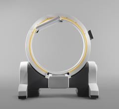
Digital medical technology company Brainlab unveiled Loop-X, which it calls the first mobile intraoperative imaging robot, at the 2019 North American Spine Society annual meeting (NASS 2019), Sept. 25-28 in Chicago.

Time September 26, 2019
arrow
News | Orthopedic Imaging

September 5, 2019 — An ahead-of-print article published in the December issue of the American Journal of Roentgenology ...

Time September 05, 2019
arrow
News | Ultrasound Imaging

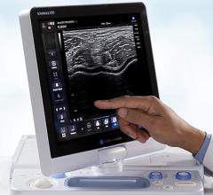
Konica Minolta Healthcare announced a new autologous biologics workshop in partnership with EmCyte Corp. The workshop is part of Konica Minolta Healthcare’s UGPro Solution, an initiative that provides complete solutions for ultrasound-guided procedures and therapies featuring the company’s Sonimage HS1 and Sonimage MX1 ultrasound systems, and including hands-on education and training.

Time June 12, 2019
arrow
Technology | Artificial Intelligence

Radiology artificial intelligence (AI) provider Aidoc announced the U.S. Food and Drug Administration (FDA) has cleared the world’s first AI solution for triage of cervical spine fractures. The latest 510(k) clearance is Aidoc’s third, coming just weeks after the FDA cleared Aidoc’s pulmonary embolism solution. By prioritizing suspected C-spine fracture cases in a radiologist’s worklist, Aidoc’s AI-powered triage automatically prioritizes critical cases, ensuring they are diagnosed by a radiologist in the most timely manner.

Time June 11, 2019
arrow
News | Ultrasound Imaging

May 29, 2019 — QT Ultrasound recently showcased its advanced ultrasound technology at the Acoustical Society of America ...

Time May 29, 2019
arrow
News | Ultrasound Imaging

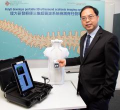
The Hong Kong Polytechnic University (PolyU) announced the development of a palm-sized 3-D ultrasound imaging system for radiation-free scoliosis assessment, dubbed Scolioscan Air. The system can bring accurate, safe and cost-efficient mass screening to schools and anywhere in the community.

Time May 03, 2019
arrow
Subscribe Now