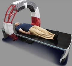
The Philips Brilliance iCT 256-slice scanner can perform a full body scan in less than a minute.
Advancements in newer 64-slice computed tomography (CT) systems and the introduction of 256- and 320-slice systems are helping to significantly reduce exposure to ionizing radiation.
In 64-slice cardiac imaging, between four and 16 images are taken and stitched together to create a full image of the heart. To create seamless images without gaps, each scan overlaps the previously scanned area by as much as 80 percent, said Michael Poon, M.D., FACC, professor of medicine and radiology, director of advanced cardiac imaging, Stony Brook University Medical Center, Stony Brook, N.Y. He said the number of images that are needed and the required overlap adds up to a lot of additional patient radiation exposure. The number of images and overlap is significantly reduced or eliminated with 256- and 320-slice systems.
The speed of the new high-slice systems and the large coverage area in each scan also help eliminate motion blur associated with pediatric patients, patients with fast heart rates or those who cannot hold their breath.
320-Slice CT
Poon said the single, large volume of the Aquilion One 320-slice system allows for a big reduction in radiation because its 16 cm imaging area can image the whole heart in one scan. This eliminates the need for stitching multiple images together and overlap imaging.
Contrast dose is also reduced with the 320-slice scanner, because only one scan is usually required, instead of several stretched out over several seconds. Poon said an average of 80-90 cc of contrast agent is used for 64-slice exams, because of the duration and number of images taken. Stony Brook Medical Center reduced this amount to as low as 50 cc with the Aquilion One.
The speed also enables scans of squirmy pediatric or neonates without the need to sedate them, Poon added.
With less radiation, lower contrast usage, fast speed, a patient weight limit of up to 600 pounds and the versatility to image any patient for nearly any condition, Poon said he can’t think of any downside to the technology, other than its price.
256-Slice CT
The Brilliance iCT 256-slice scanner can perform a full body scan in less than a minute, and Philips says it exposes patients to 80 percent less radiation than traditional 64-slice CT machines.
The University of Chicago Medical Center’s older 64-slice scanners use retrospective gating. This technique keeps the X-ray source on for the entire scan, and ECG data is used to select the best images to stitch together. However, “You get a whopping dose,” said Michael Vannier, M.D., FACR, professor of medicine at the University of Chicago Medical Center.
The center purchased a Brilliance iCT, which images a larger volume, so only about two images are needed for stitching and there is much less image overlap. The gantry speed is also much faster, helping to reduce motion artifacts. “The doses are reduced dramatically,” Vannier said.
He explained the system also has a setting for 100 kV, which helps lower the dose. The 64-slice systems only have settings for 120 and 140 kV.
Lowering 64-Slice Dose
The GE Healthcare CT750 HD scanner is designed to help reduce dose by up to 83 percent for cardiac scans. That claim was confirmed by James Min, M.D., director of the cardiovascular CT lab at New York Presbyterian Hospital and assistant professor of radiology at Weill Cornell Medical College. He said the system helped lower the average dose among his cardiac patients from an average of 5.5 millisieverts (mSv) to an average of 1.2 mSv. This includes all types of patients. Forty percent of these patients, who have a smaller body size, receive doses of less than 1 mSv.
“I think the scanner offers better image quality and increased patient safety,” Min said.
New York Presbyterian will likely purchase a second high-definition CT system later this year for its women’s health center. Min said the system will help promote a lower radiation dose in female patients.


 August 09, 2024


