Editor's Note: This article is an introduction to the breast biopsy systems comparison chart that ran in the September 2010 issue of Imaging Technology News. The chart can be found under the comparison chart tab at the top of the page.
Biopsies were once the domain of surgeons, but with the introduction of image-guided needle biopsies in 1992, radiologists have increasingly taken over that diagnostic role.
During the past decade, there was a continuing trend away from invasive approaches and non-image-guided approaches in favor of percutaneous needle biopsy (PNB) and imaging-guided percutaneous biopsy (IGPB).3 Consequently, radiologists are performing an increasing share of biopsies across all anatomic regions.
Today, the transition to needle biopsies directed by ultrasound or stereotactic mammography has made the use of surgical biopsies very rare. The minimally invasive biopsy process reduces deformity and tissue trauma. It is also faster and more cost-effective, since it is done in an outpatient setting.
The radiologists’ role in breast biopsies has gotten progressively stronger in recent years, since at least 86 percent are performed using imaging guidance.1 Between 1999 and 2004, the breast biopsy utilization rate per 100,000 Medicare beneficiaries increased by 43 percent, and in 2004, a total of 144,697 breast biopsies were performed. From 1999 to 2004, procedure volume increased by 101 percent among radiologists, compared with 32 percent among surgeons. By 2004, radiologists performed 63 percent of all breast biopsies, compared with 32 percent by surgeons and 5 percent by others.1
Minimally Invasive Technologies
Minimally invasive breast biopsy procedures, such as vacuum-assisted breast biopsy (VAB), core needle biopsy (CNB) and fine needle aspiration (FNA) are expected to grow in the coming years, causing the volume of image-guided breast biopsies to rise.2 Needles are guided by stereotactic, magnetic resonance imaging (MRI) and ultrasound.
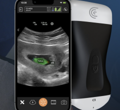
Ultrasonography-guided core needle biopsies are the modality of choice. Following administration of a local anesthetic, a 14- or 18-gauge, spring-loaded core biopsy needle is inserted during real-time imaging with the transducer.
Ultrasound-guided fine-needle aspiration is another option when a core biopsy cannot be performed because the lesion is located adjacent to sensitive structures, such as implants for the pectoralis muscle. Fine-needle aspiration is also used to evaluate complicated breast cysts and lymph nodes. Drawbacks of fine-needle aspiration (relative to larger core needle biopsy) are that it is limited to cytologic, not histologic, examination and that it yields a higher false-negative rate.4
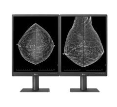
Stereotactic mammography biopsies are guided using a pair of digital mammogram images generated by a specialized, low-dose X-ray machine. The system pinpoints the exact location of a mass by using a computer and X-rays taken from two different angles.
A stereotactic core biopsy is usually performed when the lesion is calcified or on masses that are visible only on mammography. The biopsy needle used for this procedure is vacuum-assisted.
This method has some limitations that ultrasonography-guided biopsy does not. The patient must be able to get on the system’s table and hold a prone position for the duration of the 45-minute procedure. Lesion position can also prevent accessibility if it cannot be reached through the hole in the table where the breast is positioned. Most tables also have a weight limit of 300 pounds, limiting use for bariatric patients.4
The MRI-guided breast biopsy vacuum-assisted technique is used if a lesion cannot be identified, or is not visible, on ultrasound. The technique has the advantage of not using ionizing radiation, as in stereotactic biopsies, but the technique is expensive.
Fine needle aspiration uses a thin 18- to 23-gauge needle. Biopsy sites are selected for and guided by radiologists with fluoroscopy, computed tomography (CT) or MRI.
References:
1. Levin D.C. “Current practice patterns and recent trends in breast biopsy among radiologists and surgeons.” Journal of the American College of Radiology. 2006 Sept. 3 (9):707-9.
2. “Global Markets for Breast Biopsy Procedures 2006 report.” Research and Markets, 2006, www.researchandmarkets.com
3. Sharon W. Kwan. “Effect of Advanced Imaging Technology on How Biopsies Are Done and Who Does Them.” Radiology, June 29, 2010, 10.1148/radiol.10092130
4. Alice Rim, et al. “Trends in breast cancer screening and diagnosis.” Cleveland Clinic Journal of Medicine, March 2008, vol. 75, supplement 1


 July 29, 2024


