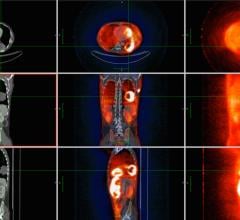
April 28, 2010 - Molecular imaging can help physicians identify aortic dissection and help guide treatment, according to research published in the May issue of The Journal of Nuclear Medicine.
Dissection occurs when a tear in the wall of the aorta causes blood to flow between the layers of the wall of the aorta and force the layers apart. Aortic dissection is the tenth leading cause of death in Western societies. It is the second most frequent cause of acute chest pain, but it is difficult to identify. However, positron emission tomography (PET) with the imaging agent fluorodeoxyglucose (FDG) and computed tomography (CT) may help determine the age of an aortic dissection, the degree of risk and the need for surgery.
"Many conventional forms of imaging are not able to clearly differentiate between acute and chronic dissection," said Hans-Henning Eckstein, M.D., Ph.D., professor at the Technical University of Munich in Germany and the corresponding author of "Imaging of Acute and Chronic Aortic Dissection by 18F-FDG PET/CT."
He added, "It is critical to patients' survival that doctors are able to verify acute or exclude chronic aortic dissection so they can decide the best course of treatment. [These might be] rushing the patient to surgery in some cases or using beta blockers to lower the blood pressure."
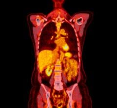
In the Munich study, researchers examined patients who had symptoms of aortic dissection and patients with chronic asymptomatic dissection using FDG PET/CT to acquire images of the affected area, just above the heart. These images were studied to determine the difference between the two forms of aortic dissection. The researchers reported that acute dissection of the aortic wall led to elevated metabolic activity in fresh lacerated segments of the aortic wall, while stable chronic aortic dissection showed no increased metabolic activity.
Researchers speculate that increased metabolic activity in cases of acute aortic dissection is due to repair mechanisms of the aortic wall injury, causing cell activation and accumulation. The low metabolic activity in chronic aortic dissection is due to scar tissue. The searchers concluded further studies are needed to prove these hypotheses.
Reference: Authors of, "Imaging of Acute and Chronic Aortic Dissection by 18F-FDG PET/CT," include: Christian Reeps, Jaroslav Pelisek, Manuela Gurdan, Alexander Zimmermann, Stefan Ockert, and Hans-Henning Eckstein, of the Clinic for Vascular Surgery, Klinikum-rechts-der-Isar, Technische Universität München, Munich, Germany; Ralph A. Bundschuh and Markus Essler, Clinic for Nuclear Medicine, Klinikum-rechts-der-Isar, Technische Universität München, Munich, Germany; and Martin Dobritz, Institute for Radiology, Klinikum-rechts-der-Isar, Technische Universität München, Munich, Germany.
For more information: www.snm.org


 July 02, 2024
