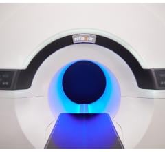
November 3, 2009 - Philips unveiled major advancements in its Brilliance CT Big Bore platform inlcuding its RapidView 4D Reconstruction System, Amplitude Binning Algorithm, and Metal Artifact Reduction, at the 51st Annual Meeting of the American Society for Radiation Oncology held from Nov. 1-5, in Chicago, Ill.
This enhanced oncology platform allows for increased accuracy in lesion localization, more efficient image reconstruction and improved workflow across patient marking and treatment.
New enhancements to Philips Brilliance CT Big Bore Oncology configurations help to improve workflow through a 4D visualization and reconstruction system. Ideal for patient positioning, this system is designed to decrease the time between imaging and patient treatment, without sacrificing image quality.
The key benefits and features include:
• RapidView 4D Reconstruction System: An 85-cm bore and true 60-cm scan field of view make 4D imaging possible, ensuring the highest quality and accuracy of images. In addition, by reducing the time it takes for scanned images to be reconstructed, this system helps clinicians to speed up motion evaluation and initiate treatment.
• Amplitude Binning Algorithm: Through 4D correlated imaging, this system allows patients to breathe freely while capturing and sorting images according to inspiration/expiration data points along a respiratory waveform. This is especially beneficial for patients that have difficulty holding their breath due to tumors in the lungs.
• Metal Artifact Reduction: This technology helps clinicians view critical structures near the sites of density metal objects or other metal in the body, and helps enable them to more accurately visualize tumors.
• “Sim Enhancement” Blocking Technology: This technology makes it possible for clinicians to “mark” an area for treatment and then initiate treatment via one workstation. Specifically, radiation therapists are able to send data directly the CT console for treatment.
• Tumor L.O.C. (or Localization On Console): This exclusive software enables the clinician to run a simple simulation at the scanner console and then quickly and accurately localize the tumor and “mark” the patient. Features include:
• Import of multiple phase datasets as well as a routine CT
• Contour on any phase and apply it to a chosen primary phase
• Dynamic DRR/DCR
• Dynamic MPR & Axial
• Maximum, minimum and average intensity projection dataset generation
Philips is also showing its third-generation Time-of-Flight technology for the GEMINI TF PET/CT and GEMINI TF Big Bore PET/CT platforms. Additional solutions from Philips CT, Nuclear Medicine and Radiation Oncology will also be on display, including:
• Tumor Tracking application
• PET Application Suite 2.0
• Pinnacle³ Version 9 Radiation Treatment Planning Software
• SmartArc technology
• SmartEnterprise data management technology
For more information: www.philips.com/healthcare


 August 09, 2024


