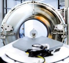
August 3, 2009 - A new lung cancer detection device, the Lung Cell Evaluation Device (LuCED), uses an automated 3D cell imaging platform aimed at detection of pre-cancerous and cancerous cells in sputum.
The technology was developed by researchers at VisionGate, Inc., in collaboration with scientists from the University of Washington. Michael Meyer, M.S., will present a study on LuCED on Tuesday, August, 4, 2009, at the 13th World Conference on Lung Cancer (WCLC), organized by the International Association for the Study of Lung Cancer (IASLC) in San Francisco, Calif.
In this study, the investigators assessed a large number of normal cells and cancer cells. LuCED automatically produces 3D cell volumetric data, allowing for the measurement of 3D cellular features that correlate with abnormal conditions. The cell measurements are then translated into an analytical score that reflects the patient’s cancer risk. LuCED, based on 3D cell analysis, produced near perfect discrimination between normal and cancer cell morphology.
“Based on abnormal cell prevalence counts in sputum from patients with cancer, we estimate that the LuCED test demonstrates near-perfect specificity (no false positives) while maintaining sensitivity that exceeds 90 percent for patients with lung cancer cells in their sputum,” says Michael Meyer, M.S, lead author and vice president for image engineering at VisionGate. “This type of 3D analysis provides an unobstructed and unambiguous representation of normal and abnormal cell morphology, making the LuCED test an effective means to guide the physicians’ further diagnostic workup, including diagnostic CT or bronchoscopy.”
For more information: www.spectrumscience.com


 April 18, 2025


