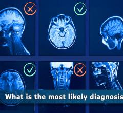
May 6, 2009 - Researchers at Dartmouth-Hitchcock Medical Center found a correlation among tumor aggressiveness, MRI-enhancement and intraoperative fluorescence in a study in which they investigated the use of a new florescence-guided technique during glioma resection surgery.
The results of this study, Fluorescence-Guided Tumor Resection: Correlation between Local Fluorescence and MRI-Enhancement, were presented by David W. Roberts, M.D., on May 5, 2009, during the 77th Annual Meeting of the American Association of Neurological Surgeons in San Diego. Co-authors are Kathryn Fontaine, BS, Brent T. Harris, MD, Alexander Hartov, PhD, Frederic Leblond, PhD, S. Scott Lollis, MD, Keith D. Paulsen, PhD, and Pablo Valdes, BA.
"Gliomas can present a challenge for surgical resection (removal) because they invade normal brain tissue that may be highly functional, so it is crucial to develop techniques for improved visualization of the tumor's margins. Surgical removal of a brain tumor can be aided using a new technique in which tumor tissue is fluoresced during surgery," stated Dr. Roberts. Early studies of this technique by a small number of institutions and a recent European multicenter trial have been reported.
This investigation was undertaken to better understand what tissue was actually fluorescing and to compare the tumor tissue's intraoperative appearance with that on preoperative imaging studies such as MRI. Twenty patients undergoing resection of glial tumors (a mix of low-grade and high-grade) were subjects for this investigation.
Patients were administered a 20mg/kg dose of 5-ALA (5-aminolevulinic acid) three hours prior to surgery. During the operation, a modified operating microscope capable of illuminating the surgical field in a special blue light was utilized. Under this condition, some tumor tissue fluoresces to a vivid pink color, while normal brain does not. A computer-based navigational system was used during surgery to identify the exact locations of tissue that fluoresced (or did not) and biopsy specimens were taken from these multiple sites. Knowing the location of each tissue sample enabled correlation of the intraoperative appearance with that of the tissue's appearance on preoperative MRI. The following clinical results were observed:
- Slower growing tumors, which are well recognized as generally showing little contrast-enhancement on MRI, demonstrated little fluorescence.
- Highly malignant tumors usually showed areas of contrast-enhancement as well as areas of non-enhancement on MRI, and at surgery the areas of fluorescence correlated very highly with MRI-enhancement.
- Tumors that were predominantly slower growing and non-enhancing but harboring small areas of enhancement demonstrated corresponding areas of fluorescence during surgery.
- Two abnormalities that enhanced on MRI but proved to not be tumors on pathological examination did not show fluorescence during surgery.
- No normal brain tissue fluoresced.
Demonstration of a high correlation among tumor aggressiveness, MRI-enhancement and intraoperative fluorescence is of considerable help in understanding the value of this new fluorescence technique.
"Although the operating microscope and navigational systems are both of great benefit during brain tumor resection, it can be very difficult for a surgeon to visually distinguish tumor tissue from normal brain. This new technique "color codes" tumor to be removed, and shows great promise for enabling more complete resection of a tumor as well as preservation of surrounding normal tissue and critical brain functions," concluded Dr. Roberts.
Funding for this research was provided by the National Institutes of Health (NIH). The author reports no conflicts of interest.
Founded in 1931 as the Harvey Cushing Society, the American Association of Neurological Surgeons (AANS) is a scientific and educational association with more than 7,400 members worldwide.
Source: American Association of Neurological Surgeons
For more information: www.aans.org
 July 25, 2024