March 4, 2009 - Physicians will be able to move away from pure surgical approaches and utilize less invasive techniques for the treatment of structural heart disease with the help of cardiovascular ultrasound technology, according to a new guideline document in the March issue of the Journal of the American Society of Echocardiography (JASE).
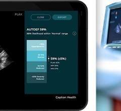
The use of echocardiography, or heart ultrasound, to guide a physician through a procedure as it is being performed has allowed physicians to treat some patients with complex conditions without open heart surgery, and has made the minimally invasive procedures to correct the conditions safer and faster, while improving the overall results for many patients.
Heart Ultrasound has been used for decades to diagnose heart conditions and is highly regarded by physicians world-wide for its ability to produce live, moving images of the heart as it works, for it’s portability, and because it is non-invasive and radiation-free, making it appropriate for use on patients of all age groups. Because echocardiography provides a safe and real-time view of the cardiovascular system, physicians can now use echocardiography to guide minimally invasive procedures in the heart catheterization and arrhythmia laboratories for conditions that once required open heart surgery.
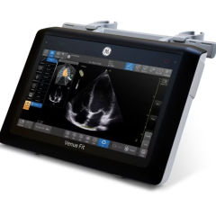
“A major advantage of echocardiography over other advanced imaging is that echocardiography is mobile and real time. Echocardiograms can be recorded at the bedside, in the cardiovascular intensive care unit, in the emergency room—virtually any place that can accommodate a wheeled cart or even a machine the size of a laptop,” said Richard E. Kerber, M.D., a lead author of the article. “This tremendous advantage allows for the performance of imaging immediately before, during, and after various procedures involving interventions.”
The guideline document outlines each type of echocardiography study as it relates to specific procedures, and makes recommendations to help guide the selection of the "right" type of echocardiogram for the procedure being performed. Procedures addressed in the document include: transatrial septal catheterization , using balloons to open tight mitral valves, closing holes in the heart (atrial septal defects)with clamshell devices, guiding the placement of artificial valves to replace degenerated native valves and more.
The ASE Guideline Document, “Echocardiography-Guided Interventions” discusses all modalities of echocardiography, but emphasizes "intracardiac echocardiography" or ICE, which is a newer type of echo that places the imaging probe within the heart during these procedures.
“When working on the heart with these less invasive techniques, imaging the procedure as it is being performed (in real time) with echocardiography has taken on a central role in selecting the right patients to work on, guiding the procedures as they are being performed, and monitoring their safety during the procedure,” said Dr. Frank Silvestry, one of the lead authors of the guideline document. “The use of imaging techniques to guide even well-established procedures enhances the efficiency and safety of these procedures.”
This is the first document of its kind that sets the standard for imaging during procedures, and serves as an important reference to a wide variety of physicians. View the full article at www.asecho.org
The American Society of Echocardiography (ASE) is a professional organization of physicians, cardiac sonographers, nurses and scientists involved in echocardiography, the use of ultrasound to image the heart and cardiovascular system. The organization was founded in 1975 and has more than 15,000 members nationally and internationally.
For more information on ASE, visit www.asecho.org or ASE’s Public Information site, www.SeeMyHeart.org


 March 06, 2024


