December 12, 2008 - Using a three-dimensional (3D) image-guided system to help place screws in the spines of patients results in safe and accurate surgery with a decrease in the number of misplaced screws, and subsequent injuries, seen in more traditional operations, say neurosurgeons at Mayo Clinic in Florida.
In the December 9, 2008 online edition of the Journal of Neurosurgery: Spine, Mayo physicians published the largest study yet using 3D image-guided technology to place screws in the spine for spinal fusion procedures. The screws are used to stabilize the spine in patients who suffer from collapsed discs or compressed nerves.
Specifically, after implanting 1084 “pedicle” screws in 220 patients, surgeons reported a nerve injury rate of less than 1 percent. Additionally, less than 1 percent of the screws in this study were considered to be significantly misplaced. That compares to a reported nerve injury rate of up to 8 percent and a misplacement rate of up to 55 percent using standard technology. As well, re-operation for removal of a misplaced screw has been reported in other surgical literature to be as high as 6.5 percent but occurred in less than half of one percent of all patients in the Mayo study, according to the researchers.
“Using 3D image-guided technology to help us place these screws results in a much better outcome for our patients,” says Mayo Clinic neurosurgeon Eric Nottmeier, M.D., the study’s lead investigator. “In addition to the decreased incidence of nerve root injury, this technology allows us to place larger screws into the spine, which can also increase the success rate of the operation.”
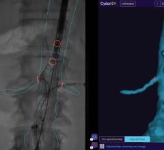
The technology uses a special camera on a computer that uses infrared light to track a surgical instrument in 3D space. The surgeon places the instrument on the patient’s spine and navigates the spine using the computer. The surgical instrument is used to determine the best entry point and trajectory for each screw. An image-guided screwdriver is used to place a screw.
In most other institutions, pedicle screws are placed using a freehand technique or by fluoroscopy, which uses X-rays to capture a one-dimensional image on a television screen of the process of screw placement. Not only is the image less detailed, but both patients and the operating room staff can be exposed to radiation and must use lead clothing for protection, Dr. Nottmeier says. Almost all patients in this study were given a CT scan following surgery so that a radiologist could independently determine how well the screws were placed.
“Every person’s spine is a little bit unique,” Dr. Nottmeier said, “and unexpected variations in bone shape and density can make screw placement in the spine more challenging, especially in patients who have had previous spine surgery.” Almost half of the patients in the Mayo study had a previous spine surgery.
“This technique allows us to have the best view possible of the vertebrae as we operate,” Dr. Nottmeier said.
Based on the success of the technique, the image guidance system is now used in all spinal screw operations at Mayo Clinic’s campus in Florida.
Two different image guided systems were used in this study: the “Stealth Treon,” manufactured by Medtronic of Littleton, Mass., and the “BrainLAB Vector Vision,” from BrainLAB in Westchester, IL. Nottmeier is a paid consultant for BrainLAB, however, this study was done independently and did not involve any company funding.
Co-authors of the study include Phillip M. Young, M.D., Department of Radiology, Mayo Clinic in Florida, and Will Seemer, B.A., Department of Chemistry, from the University of North Florida in Jacksonville.
Source: Mayo Clinic
For more information: www.mayoclinic.org/news

 February 01, 2024
