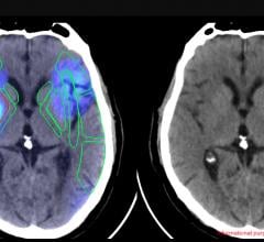
Siemens offers an integration of CAD into its advanced 3D applications, as well as the use of CAD in the PACS workflow.
The syngo Lung CAD is a computer-aided detection (CAD) software for automated detection of solid pulmonary nodules in thoracic CT studies. This CAD tool plays a valuable role as an effective second reader to aid in the interpretation of today’s complex chest CT data sets, which may contain hundreds of images. syngo Lung CAD is available on the syngo MultiModality Workplace (MMWP) for use with syngo CT Oncology. Additionally, to enable the CAD results to be displayed on PACS workstations, syngo Lung CAD is integrated in the reading workflow of syngo Imaging and syngo Imaging XS.
The syngo PE Detection is a second reader tool to help detect segmental and sub-segmental filling defects in CT Angiography (CTA) studies of the thorax. syngo PE Detection is integrated with the syngo Circulation application, enabling an efficient reading and reporting process.
The syngo CXR CAD is a second reader product for the detection of lung nodules in digital chest X-ray studies. syngo CXR CAD processes images of the posterior-anterior (PA) view and is optimized for the detection of lung nodules from 8 mm to 30 mm in size. syngo CXR CAD is validated for use with images from the Siemens AXIOM Aristos Digital X-Ray system, and the results can be displayed on PACS workstations.
The syngo MammoCAD is a computer-aided detection solution for full-field digital mammography (FFDM) and generates CAD marks to highlight suspicious areas, such as masses and microcalcifications. With syngo MammoCAD up to 40 percent of normal cases show no false positive CAD marks , saving the radiologist interpretation time during the second read phase. In combination with the syngo MammoReport workstation, additional features include:
- Breast density in MammoBrowser.
- Show More, Show Less, which customizes the display of CAD markers.
- Additional CAD information provides the number of calcifications in a cluster.
- Corridor of interest: Identification of marked lesion in second view using CAD findings.
The syngo Colonography PEV (polyp enhanced viewing) is an automated second reader tool for the visualization of lesions in the colon. When a specific PEV finding is selected, the tool automatically goes to the PEV-marked polyp in the 3D endoscopic and multi-planar reformation (MPR) views. It helps detect polyp-shaped objects that are between 6 mm and 25 mm in size. In addition, automatic size measurement tools support accurate polyp measurement. syngo Colonography PEV can be used both in clean-prepped and solid-liquid tagged protocols. The product was developed using an extensive database of more than 1,700 CT colonography cases from more than 15 clinical sites worldwide. syngo Colonography PEV is available on the syngo MultiModality Workplace (MMWP), syngo CT Workplace and with syngo Imaging.
The syngo TrueD, a whole-body hybrid imaging solution provides advanced image analysis and comparison to help make more informed diagnosis, therapy and follow-up decisions. The latest version of syngo TrueD includes numerous workflow-enhancing features, such as user-configurable layouts and new VOI trending analysis tools. Other main features are the simultaneous display of three time-points, support for respiratory gated PET•CT studies, deformable registration, and saving DICOM RT structured objects for use with therapy planning systems. syngo TrueD is available on syngo MultiModality Workplace (MMWP) and with syngo Imaging.


 June 06, 2023


