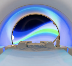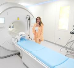
GE Healthcare says it is breaking the bonds of traditional magnetic resonance scanning with the introduction of its latest MR scanner, the Signa MR750 3.0T.
Equipped with powerful gradients, easy-to-use workflow features and the company’s advanced Thermal Management System, the Signa MR750 delivers up to 60 percent additional anatomical coverage and resolution unit per time. The system also allows for up to five times the imaging performance over previous generations, increasing the freedom for advanced application development.
Relevant clinical applications include LAVA-Flex, a dual-echo acquisition technique that raises the bar on existing sequences to provide consistent, detailed, three-dimensional abdominal images in one breathhold. In addition, LAVA-Flex produces in one scan four different contrasts, allowing the user to select the output image types - in-phase, opposed-phase, water, and fat. With this new application, clinicians can now conduct a complete liver exam in 15 minutes.
LAVA- Flex offers ‘fat only’ and ‘water only’ images in addition to high quality in-phase and opposed-phase images in a single breathhold, a sequence that has quickly become a routine part of all our abdominal sequence protocols at 3.0T, according to Elmar Merkle, M.D., professor of Radiology, Head of Body Magnetic Resonance Imaging and Medical director of the Center for Advanced Magnetic Resonance Development at Duke University.
VIBRANT- Flex is a new application that allows for fat-free breast imaging with high spatio-temporal resolution. This application catches the shortest in- and out-of phase echoes to keep scan times comparable to single echo acquisitions, even though twice the amount of data is collected.
VIBRANT Flex optimizes acquisition with a high signal-to-noise ratio (SNR) for acquiring high quality water and fat images. This capability lets the user prescribe thinner slices for high spatial resolution imaging.
PROPELLER 2.0 enables strong performance in reportedly all imaging planes with the implementation of the No Phase Wrap (NPW) technique. NPW allows virtually ghost-artifact-free, motion-immune scans in sagittal, coronal, axial and oblique planes. Since this technique effectively deals with the aliasing artifact, PROPELLER 2.0 is now more robust performing small field-of-view (FOV) scans.
With GE-exclusive 3D SWAN technique, you can capture and delineate small vessels and microbleeds as well as large vascular structures and iron and calcium depositions in the brain. 3D SWAN's unique, multi-echo acquisition technique and postprocessing algorithm provide wider tissue bandwidth, 2-4 times more SNR and more contrast in a simple to use, robust package within clinically relevant scan times. With such broad clinical properties, 3D SWAN may aid clinicians in diagnosing among others patients with ischaemic and cerebral brain diseases, arteriovenous malformations and neurodegenerative diseases.


 July 25, 2024


