September 29, 2008 - Philips is developing a system that uses ultrasound-activated microbubbles for chemotherapy drug delivery designed to increase the effectiveness and reduce the side effects.
The system proposes the use of drug-loaded microbubbles, no larger than red blood cells, which can be injected into the patient’s bloodstream, tracked via ultrasound imaging and then ruptured by a focused ultrasound pulse to release their drug payload when they reach the desired spot. Because the drugs would only be released at the site of the diseased tissue, the patient’s total body exposure to them could be limited. For certain types of treatment – for example, chemotherapy for breast cancer – this could help to reduce unpleasant side effects.
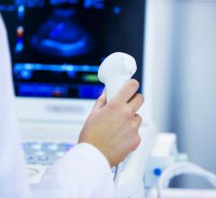
The use of microbubbles in conjunction with medical ultrasound imaging is not new. However, at the moment in clinical practice, microbubbles are only used as contrast agents for example to highlight blood in the ultrasound images – an application that relies on the fact that microbubbles reflect ultrasound much better than blood or soft tissue.
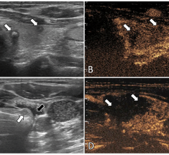
The drug delivery technology being developed by scientists at Philips Research continues to utilize the contrast-enhancing capabilities of microbubbles to help ultrasound operators to locate tumors – based on their density and the fact that tumors typically grow a recognizable network of small blood vessels around themselves. What’s new is that it then shatters the shells of the microbubbles in these blood vessels using a focused high-energy ultrasound pulse. As a result, the drugs contained in the microbubbles are released directly inside the tumor.
Philips is working with several academic partners, including the University of Virginia (USA) and the University of Muenster (Germany), to refine the technology. Clinical institutions, such as The Methodist Hospital in Houston (USA), are also actively researching this new and exciting field of ultrasound mediated drug delivery.
“More and more, patients are demanding treatment options that allow them to maintain their quality of life during the treatment regime, without sacrificing treatment efficacy,” comments King Li, MD, chair of the Department of Radiology at the Methodist Hospital in Houston (USA) and professor of Radiology, Weill Cornell Medical College (USA). ”The non-invasive nature of ultrasound mediated delivery is a step in this direction. Work at our and other institutions using ultrasound for drug delivery and treatment guidance has shown the potential of this technology in pre-clinical studies.”
For more information: www.medical.philips.com

 July 19, 2024
