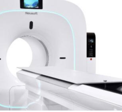
September 5, 2008 - Digirad Corp. today announced the initial clinical trial of its new SPECT imaging system incorporating new proprietary technology to correct attenuation, an inherent issue in cardiac SPECT imaging.
The new system, Cardius X-ACT, will be exhibited at the 2008 Annual Scientific Session of the American Society of Nuclear Cardiology (ASNC) Sept. 10-14, 2008, at the John B. Hynes Convention Center in Boston.
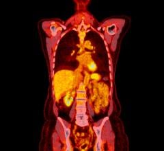
Cardiac SPECT (single photon emission computed tomography) used for myocardial perfusion imaging is a noninvasive test to assess the heart’s structure and function. Small amounts of radioactive substances are injected into the patient’s vein, and gamma cameras produce images of the blood flow in the heart. These SPECT images are used to identify blockages in coronary arteries, determine whether a patient has had a heart attack, evaluate risk of a heart attack, and assess a patient’s condition after bypass surgery or angioplasty.
However, attenuation can cause misdiagnosis of the images, which is one of the most significant issues in cardiac SPECT. There are alternative solutions that attempt to address this problem, but Digirad said they involve tradeoffs, such as noisy images, truncated scans, lengthy procedures with risk of excess movement by patients, or higher levels of radiation dosage.
Research based using the company’s Cardius 3 triple-head SPECT camera, and feedback from initial clinical trials of the Cardius X-ACT at the University of California, Los Angeles (UCLA) is promising, compared to other SPECT systems at competitive price points, Digirad said. The company said the new device shows improvement in imaging clarity and accuracy, more rapid imaging, and a significant reduction in radiation dosage.
Two other clinical studies are planned for the Cardius X-ACT system, which is expected to be available for sale sometime in mid-2009 pending FDA approval.
For more information: www.digirad.com


 July 30, 2024


