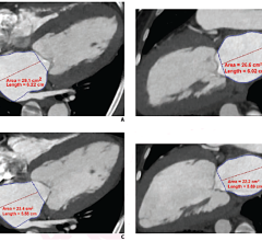
July 29, 2008 - Physicians and medical physicists often spend hours drawing lines around tumors and organs in CT images, causing a major bottleneck in cancer treatment.
A new semi-automatic user-interface could reduce the time and fatigue associated with this meticulous task.
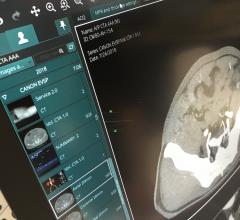
Radiation therapy begins with a CT scan in which 100 or so individual images (slices) are combined to create a 3D map of the region around the tumor. During the following segmentation step, all the organs and sensitive tissues must be identified and outlined for each slice, so that the medical physicist can plan a treatment that provides the highest dose to the target, while sparing the surrounding healthy tissue.
The resolution in CT scans is constantly increasing, which means more slices and more time required for segmentation. In addition, moving organs like the lung are starting to be scanned several times to form a time sequence. This can multiply by 10 the number of images an expert must analyze.
To help with this overwhelming load, Yu-chi Hu (huj@mskcc.org) and Gig Mageras of the Memorial Sloan-Kettering Cancer Center in New York, NY, along with Michael Grossberg of the City College of New York have developed a computer program that can segment organs with just a small amount of user input. Starting with one CT image, the user makes a crude outline of each organ. The computer takes this rough sketch and plugs it into a statistically-based algorithm, which it then uses to generate contours in subsequent images. The user checks the computer-drawn boundaries and can correct mistakes with tiny brushstrokes on the computer screen. These corrections are reincorporated by the software to better refine the algorithm.
With funding from the NIH, the team tested the user interface on several CT scans and found that on average an image could be segmented in roughly 6 seconds with the computer's help, instead of the 30 seconds or more in the unaided case. The researcher's algorithm correctly identified 98 percent of the image pixels, which was a higher precision than other contour-drawing algorithms that the researchers tested. The group is now planning to put the system into clinical use within 6 to 9 months.
For more information: www.aapm.org
Source: American Association of Physicists in Medicine

 February 01, 2024
