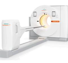
June 3, 2008 - Seattle Children's Hospital is the first pediatric facility to install a 64-slice GE Discovery VCT PET/CT, combining the system with tailored pediatric imaging protocols to allow Children�s radiologists to best meet the needs of its pediatric patients by more accurately diagnosing conditions and pinpointing treatment areas, while protecting children from excessive diagnostic radiation exposure.
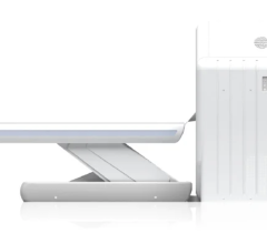
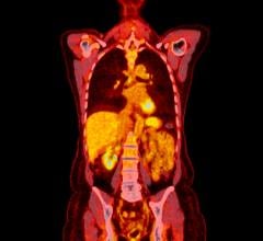
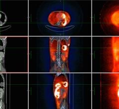
The Discovery VCT PET/CT scanner pairs Positron Emission Tomography (PET) with Computed Tomography (CT) and takes 64 pictures of the tumor site that are combined into a comprehensive 3D image allowing radiologists to determine the presence of disease, pinpoint specific biopsy sites and accurately monitor the effects of treatment.
The Discovery VCT PET/CT is most commonly used in oncology to diagnose lymphoma, neuroblastoma, brain tumors, sarcomas and thyroid cancer. Additionally, it can also assist in the diagnosis of myocardial viability, ischemia, coronary artery disease and in localizing epilepsy and seizure origin.
Diagnostic imaging with a PET/CT scanner requires a small amount of radiation. Studies have shown that these small amounts of radiation can slightly increase the risk of adverse health effects. At Seattle Children�s Hospital, PET/CT scans are designed to minimize diagnostic radiation while ensuring image quality. The scans are only performed when the benefit far outweighs the risk of adverse effects.
The PET scan is a metabolic imaging technology that detects changes in cell energy to establish whether tumors are active or inactive. The PET scan also determines if tumors have spread locally, or to outlying locations away from the primary tumor. The CT scan is then used to precisely identify the location of these cells within the childn's body.
For more information: www.gehealthcare.com, www.seattlechildrens.org


 July 02, 2024
