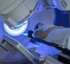
May 21, 2008 – Modern 3D computed tomography (CT) is an effective method for locating the prostatic apex for radiation therapy treatment planning in prostate cancer patients because it eliminates the need for an invasive procedure and the related side effects, according to a study in the May 1 issue of the International Journal of Radiation Oncology.
Retrograde urethrography, which involves inserting a catheter into the male urethra to inject contrast, is the standard method used to identify the area of the prostate to be treated with radiation. However, this method is invasive and uncomfortable for patients and comes with risks of side effects, such as urethral injury and infection, as well as additional costs.
Researchers at the University of Texas Health Science Center at San Antonio departments of Radiation Oncology and Urology conducted this study to determine if knowledge of the anatomic relationships of the prostatic base and the prostatic apex along with modern 3-D CT planning, could be used as a substitute for retrograde urethrography.
Fifteen patients underwent a CT simulation both with and without bladder, urethral and rectal contrast. The prostatic base and apex were identified easily and consistently on both scans by taking a side view of the patient and drawing a line from the pubic bone straight down to the floor of the pelvis. The process was repeated and confirmed in another 57 patients, leading researchers to determine that it is not necessary to subject a patient to a urinary catheter for contrast delivery.
“By using CT scans to find the prostatic apex, patients are happier and we have the same results,” said Gregory Swanson, M.D., associate professor of radiation oncology at the University of Texas Health Science Center at San Antonio and one of the study’s authors. “I stopped doing contrast in 1995 or 1996 when I realized that I knew where the prostate was without using invasive methods. I have been successful in using CT since then.”
For more information: www.astro.org


 August 09, 2024


