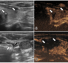
July 10, 2007 - Biomedical engineers at the University of Virginia (U.Va.) School of Engineering and Applied Science have developed a new imaging tool that hopes to dramatically improve medical ultrasounds, potentially leading to more accurate and timely diagnoses of breast cancer and other life threatening conditions.
Using Star-P software from Interactive Supercomputing, the University's biomedical engineering research team, led by Associate Professor William F. Walker, created an advanced beamforming algorithm - called the Time-domain Optimized Near-field Estimator (TONE) - which significantly improves the contrast and resolution of ultrasound images.
"The potential applications for this algorithm are almost infinite," said James H. Aylor, dean of U.Va.'s School of Engineering and Applied Science. "Not only can it be used in the medical community to benefit patients nationwide, it will have applications in the fields of radio astronomy, seismology and more."
While conventional beamforming algorithms have been used in ultrasound scanners for nearly a half century, they typically result in degraded images that are blurry or cluttered. The culprit: off-axis signals, or the sound wave reflections coming from undesired locations within the organ or tissue.
The TONE algorithm reduces undesired off-axis signals, resulting in much higher definition images, but at the price of a much greater computational load. The algorithm developed on desktop computers overwhelmed the computer's processing ability. The team solved this problem by automatically parallelizing their algorithms with Star-P to run on a powerful, memory-rich IBM Linux cluster.
For more information: www.seas.virginia.edu, www.interactivesupercomputing.com

 July 19, 2024
