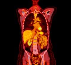
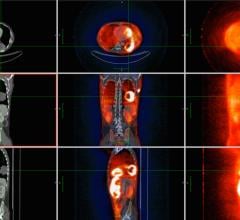
June 5, 2007 - Moving from computer simulation to patient images, researchers are now demonstrating the benefits that time-of-flight (TOF)/PET imaging can provide for cancer patients. The result? Superior images and shorter patient scan times for starters, according to a study released at the 54th Annual Meeting of SNM, the world’s largest society for molecular imaging and nuclear medicine professionals, June 2–6 in Washington, D.C.
“Our TOF/PET patient images exhibit superior image quality and suggest that shorter patient scan times could be performed in many cases,” said Amy Perkins, a Philips Medical Systems clinical site scientist based at the University of Pennsylvania in Philadelphia. “Previously, we have studied TOF/PET with computer simulations and controlled experiments to approximate the behavior within the human body, showing that we can get very good image quality with shorter scanning times,” she said. “We have now moved our investigation to clinical studies—using PET scans from patients with a wide range of body weight, with different types of cancer and with different size cancer tumors—to determine whether the scan time may be reduced significantly without sacrificing clinical content,” added Perkins. “In our study, we are getting an excellent representation of what’s going on in the body,” she added.
For more information, visit: http://interactive.snm.org


 July 02, 2024
