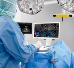
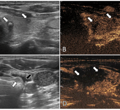
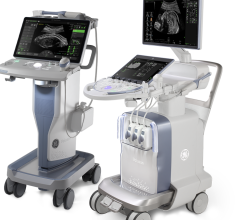
GE Healthcare highlighted at the American Heart Association (AHA) in Chicago new 4-D imaging capabilities in its Vivid 7 Dimension cardiovascular ultrasound platform, which are designed to help clinicians assess cardiovascular anatomy and LV function with more accuracy.
The Vivid 7 Dimension ‘06 system is a PC-based, software, raw data ultrasound platform engineered to deliver real-time, un-gated, un-spliced 4-D imaging and real-time 4-D full volume imaging, reportedly enabling clinicians to render 4-D cardiovascular imaging during day-to-day clinical exams. New to the Vivid 7 Dimension is real-time 4-D color flow full volume imaging, assisting clinicians in their assessment of hemodynamic information in color and real time in the same heart cycle. Also new is 4-D LV Volume – an EchoPAC Dimension option, enabling users to quantify volume information from real-time full volume Vivid 7 data. Packaged in the application is TomTec Imaging Systems technology that reportedly can provide a full assessment of the left ventricle volumes free of geometric assumptions.
The Vivid 7 Dimension ‘06 innovations also include Advance Tissue Synchronization Imaging (TSI). Time-to-peak data is derived from TSI’s “red light/green light” visualizations to quantify left ventricle synchronicity for heart failure patients or those undergoing cardiac resynchronization therapy (CRT). Also new is Automated Function Imaging (AFI), a tool that allows clinicians to assess and quantify left ventricle wall motion.
© Copyright Wainscot Media. All Rights Reserved.
Subscribe Now

 July 19, 2024
