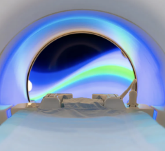
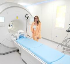
GE Healthcare has introduced the Signa HDxt as the next-generation High Definition MR scanner, available in both 1.5T and 3.0T field strengths. Highlighting the new system are two new marquee applications called Cube and IDEAL.
Available for both 1.5T and the 3.0T systems, the GE-exclusive MR imaging application, Cube, can reportedly replace standard 2D acquisitions acquired in multiple planes with a single highly-accelerated 3D volume scan. Users are able to review high-definition, 3D data from one acquisition in any plane– axial, sagittal, coronal, oblique – with no gaps or resolution loss. The submillimeter isotropic voxel size and enhanced tissue contrast typical for Cube acquisitions is intended to allow radiologists to visualize lesions as small as two millimeters and get consistent image quality and reproducible results with automated protocols optimized for each anatomy.
Also new on the Signa HDxt platform is IDEAL, an imaging technique resulting in four images, all from one acquisition. Even with challenging anatomy or metal implants, IDEAL is said to provide robust water-only images while also providing users with important information from fat images as well.
The uses of IDEAL include: uniform fat suppression, multiple contrasts and minimal artifacts.
Additional applications available for the Signa HDxt 1.5T and 3.0T MR systems have also expanded imaging visualization for neurological, vascular, cardiac, and liver diagnostic assessment. These applications include:
∑ AngioCard - Reportedly expedites vascular reporting and enhances communication with schematics, images and movies provided to reports for referring physicians.
∑ StarMap - Assesses iron deposition in the heart and liver with an automated scanning sequence, calculates T2*-R2* pixel by pixel within an region of Interest and provides decay curve, grayscale and color map displays
∑ Flow Analysis - Is said to quantify flow characteristics, which complements phase contrast, speeds Region of interest placement with Automated and Propagated trace and provides Graphic and numeric display of flow and velocity data.


 July 25, 2024


