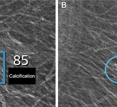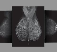
July 27, 2012 — The Journal of Ultrasound in Medicine has published a pilot study conducted at Thomas Jefferson University in Philadelphia, with funding provided by Toshiba, that explores the effectiveness of Toshiba America Medical Systems Inc.’s MicroPure image processing technique in visualizing breast microcalcifications compared with grayscale ultrasound imaging. According to the pilot study, the MicroPure software significantly improved identification of microcalcifications in the breast when compared with grayscale ultrasound imaging.
“MicroPure may have the potential to assist in the early detection of small specks of mineral deposits (calcium) associated with extra cell activity, allowing physicians to monitor changes,” said Flemming Forsberg, Ph.D., professor of radiology, Thomas Jefferson University. “The results of the study are particularly important, as they show the potential to utilize ultrasound imaging in identifying breast microcalcifications, which is less expensive and more comfortable for women.”
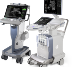
The study included women with breast calcifications, originally identified by mammography, who were then evaluated using MicroPure software on the Aplio XG ultrasound system compared with grayscale ultrasound imaging. Toshiba’s MicroPure enabled physicians to more easily visualize microcalcifications using ultrasound.
“This study is an example of Toshiba’s commitment in developing the latest diagnostic imaging technology to improve diagnoses,” said Tomohiro Hasegawa, director, ultrasound business unit, Toshiba. “As a result of this study, Thomas Jefferson University demonstrated the potential of MicroPure’s ability to visualize microcalcifications that were not as visible in gray scale ultrasound imaging.”
For more information: www.medical.toshiba.com, www.jultrasoundmed.org


 July 29, 2024

