How IQon Spectral CT is Ushering in a New Era of Medicine

Spectral CT is the next step on an evolutionary path that diverged 20 years ago with multi-slice imaging. Multi-slice was faster — and more accurate. But conventional CT scans often produce ambiguous data that can require additional testing. Rather than building scanners with ever more slices, Philips chose to unlock information in the spectrum of X-rays emitted during a CT exam.
Today its premium scanner, the IQon Spectral CT, provides on-demand spectral results through its proprietary detection-based solution. The IQon Spectral CT reconstructs data by capturing high and low X-ray energies at the same time and in the same space. And it can break down energy-specific information into slices that highlight specific materials contained in tissues.
Additionally, the IQon Spectral CT is equipped with the Spectral Diagnostic Suite, which includes Philips’ Spectral Magic Glass and the Magic Glass on PACS App. While Spectral Magic Glass allows several different spectral results to be viewed and compared at the same time for a specific region of interest, the Magic Glass on PACS App provides the ability to review and analyze spectral results from anywhere in the organization via your PACS.
“There’s a clear advantage to making the right diagnosis in the first scan,” said Sondra Miller, vice president CT global product marketing. “With the IQon Spectral CT, the first exam is the right exam. It can provide better patient experiences. And it can empower clinicians to improve clinical outcomes — while at the same time achieving the economic objectives of their organizations.”
Boosting Efficiency
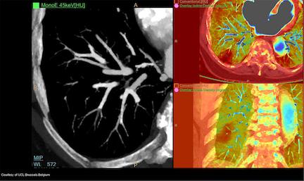
At Université catholique de Louvain (UCL) in Brussels, Belgium, an emergency patient complaining of chest pain and shortness of breath was scanned on the IQon Spectral CT. Clinicians could not clearly identify problematic pathology on conventional CT scans alone. However, utilizing spectral images, which are available on-demand on the IQon, clinicians were able to identify a perfusion defect in the right lower lung using iodine overlay results. They were then able to re-evaluate conventional scans to locate a small right lower lung pulmonary embolism (PE).
If the IQon Spectral CT had not been available retrospectively, more testing might have been necessary to confirm the perfusion defect and find the PE.
Spectral CT also expands the range of patients who can be scanned. Patients with metal implants, for example, may be scanned with minimal artifacts by viewing the data at the higher energy end of the CT spectrum. Images at high effective x-ray energies, ranging from 100 to 200 keV, are less susceptible to metal artifacts, Miller noted.
Such selective reconstructions, rendered with quantitative accuracy, are possible thanks to Philips’ unique NanoPanel Prism detector. The detector simultaneously records the energies of photons across the x-ray spectrum. Its top layer picks up the lower energies; its bottom layer records the higher ones.
Because data across the spectrum are acquired simultaneously, IQon Spectral CT requires no special preplanning or additional scans. Scan speed — and, therefore, patient throughput — are unaffected. IQon Spectral CT uses the same iPatient workflow as do most other conventional Philips CT scanners, Miller noted.
“Spectral results are always on and can always be reviewed and analyzed on-demand on PACS, anytime, anywhere,” Miller said. “This complete integration of spectral data from the acquisition through analysis — with no impact on workflow — is the key enabler for spectral CT to become the standard of care.”
Limiting Dose
X-ray dose may be reduced between 60 and 80 percent by Philips’ iterative model reconstruction (IMR) software. With IMR, IQon Spectral CT administers patient doses identical to those of Philips’ conventional high-performance scanner, the iCT.
Although IQon Spectral CT is the flagship of Philips’ CT portfolio, the premium scanner is being installed at medical institutions of all sizes, not just big teaching hospitals. Miller notes that providers all want to improve the quality of patient care and clinical outcomes. And the coming era of value-based medicine will require them to make these improvements, while keeping a lid on costs. IQon Spectral CT’s ability to support quick, confident, and cost-effective diagnoses plays into the strengths of value-based medicine, she said.
“The world of healthcare is demanding solutions that will enhance diagnostic certainty and first time right imaging,” Miller said. “This is the value proposition of the IQon Spectral CT — to provide better patient experience, enhance clinical care and, ultimately, help achieve economic objectives.”
Phillips' Disclaimer
In clinical practice, the use of IMR may reduce CT patient dose depending on the clinical task, patient size, anatomical location, and clinical practice. Consultation with a radiologist and a physicist should be made to determine the appropriate dose to obtain diagnostic image quality for the particular clinical task. Low-contrast detectability and image noise was assessed for 0.3% contrast using a Reference Abdomen Protocol — 10 mGy, 0.8/0.4 mm, Standard filter B as compared to 2 mGy IMR Smooth, L3; for 1.4% contrast using a Factory Abdomen Protocol — 10 mGy, 0.8/0.4 mm, Standard filter C as compared to 4 mGy IMR clinical indication Sharp, L3; tested on the MITA CT IQ phantom (CCTT183, Phantom Laboratories).


 December 10, 2025


