
Greg Freiherr has reported on developments in radiology since 1983. He runs the consulting service, The Freiherr Group.
BLOG: How Advanced X-Ray Systems Help Meet Outpatient Challenges

The KDR-UA shows positioning for an ankle radiograph. (Photo courtesy of Konica Minolta.)
The pressure to be efficient and effective when imaging outpatients may be rising due to imaging’s increased presence outside hospital settings. The challenges that accompany this trend are exemplified at busy orthopedic clinics, such as the Tallahassee Orthopedic Clinic (TOC) in Tallahassee, Fla.
“We try to get our patients in and out of X-ray as quick as possible so that doctors can make their plans to take care of them,” said Shelby Morris, TOC’s radiology supervisor.
Underscoring the importance of fast and effective outpatient imaging is a trend toward imaging in outpatient clinics. The Advisory Board reported in an executive research briefing December 12, 2017 that new payer and patient preferences are increasing pressure to place imaging services in freestanding facilities. This could potentially accelerate a move in imaging services from inpatient to outpatient settings, one that has long been going on, according to the Advisory Board.
Putting Patients First
Patients want answers, but they also want to be comfortable, as evidenced by a growing interest in patient concerns on the part of the imaging community.
“We try to make our patients as comfortable as possible while we’re doing the exams,” said Morris.
TOC operates two Konica Minolta U arms: one at a central location in Tallahassee, which houses 27 physicians; the other at a satellite clinic. Together they provide patient-centered orthopedic care and sports medicine to North Florida and South Georgia, according to the TOC website.
At TOC, as in other outpatient settings, space is at a premium. The highly functional Konica Minolta U-Arms feature a compact footprint that helps TOC make the most of available space.
The floor-mounted U-arms not only optimize available space, they operate efficiently and effectively, thanks in part to onboard software that allows technologists to set up procedures before patients come into the room. “Say you want an upright knee — you click on a remote and it takes the machine right to where you need it to be,” said Morris who speaks from experience with the device.
Efficiency and patient comfort were high on the list of design requirements that went into the U-arm, according to Guillermo Sander, director of product management at Konica Minolta Healthcare. “We needed to make it very, very quick and as easy on the patient as possible,” Sander said. “Patient comfort was our main driver.”
Its flexibility can spare the patient from physically moving into different positions, even when in a wheelchair, according to Morris. “You can put the detector low enough to set up like a tabletop — have patients stay in their chairs and just prop their ankles up on the detector,” she said. “The image is taken. And the patient never has to get out of the wheelchair.”
Efficiency Matters
Released commercially two years ago, the floor-mounted system KDR (Konica Digital Radiography) Advanced U-Arm swivels and tilts automatically into position for the exam chosen by the technologist. Image quality is achieved using a state-of-the-art cesium iodide digital detector that delivers high-quality bone and soft tissue images. According to specifications for the latest version of the KDR-AU, the system can move 135 degrees and travel 39 inches vertically.
This flexibility is critically important for effective, efficient and patient-friendly operation. The U-arm can image head, neck, arms and legs, as well as joints from elbows to knees to toes for ambulatory patients as well as those in wheelchairs. The physical construction of the system supports its flexibility, as does the software that guides the machine through exams.
“For the same body part, we have two or three different codes that the doctor will order,” said Sander. This allows images to be acquired so they look the way the physician wants, he added.
Related Content
May 20, 2024 — Exo (pronounced “echo”), a medical imaging software and devices company, announced the release of Exo ...
 May 20, 2024
News | Cardiac Imaging
May 17, 2024 — The Cum Laude Award-Winning Online Poster presented during the 124th ARRS Annual Meeting found that the ...
May 17, 2024 
News | Radiology Business
May 14, 2024 — University Hospitals (UH) and Siemens Healthineers announce a 10-year strategic alliance that builds on ...
May 14, 2024 
News | RSNA
May 7, 2024 — The Radiological Society of North America (RSNA) and the Radiological and Diagnostic Imaging Society of ...
May 07, 2024 
May 7, 2024 — The Magna Cum Laude Award-Winning Online Poster presented during the 124th ARRS Annual Meeting showed a ...
May 07, 2024 
News | ARRS
May 7, 2024 — The American Roentgen Ray Society (ARRS) announced that Philip Costello, MD, the 118th ARRS President and ...
May 07, 2024 
May 6, 2024 — Hvidovre Hospital has the world's first prototype of a sensor capable of detecting errors in MRI scans ...
May 06, 2024 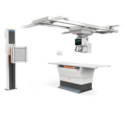
Feature | Digital Radiography (DR) | By Melinda Taschetta-Millane
Digital radiography (DR) continues to advance at a rapid pace with today’s technological innovations and evolving ...
May 06, 2024 
Feature | Radiology Business | By Melinda Taschetta-Millane
One on One interviews with radiology trailblazers and historic FDA clearances made the top-read list for April. Take a ...
May 03, 2024 
Feature | Radiation Dose Management | By Christine Book
Advances in the growing radiation dose management market are continually helping those who administer treatment to focus ...
May 03, 2024 © Copyright Wainscot Media. All Rights Reserved.
Subscribe Now